 SEPS
Society for Evolutionary Parasite Systematics
SEPSM. R. Dwyer
Research and technical report. No validated end-to-end identification performance claimed.
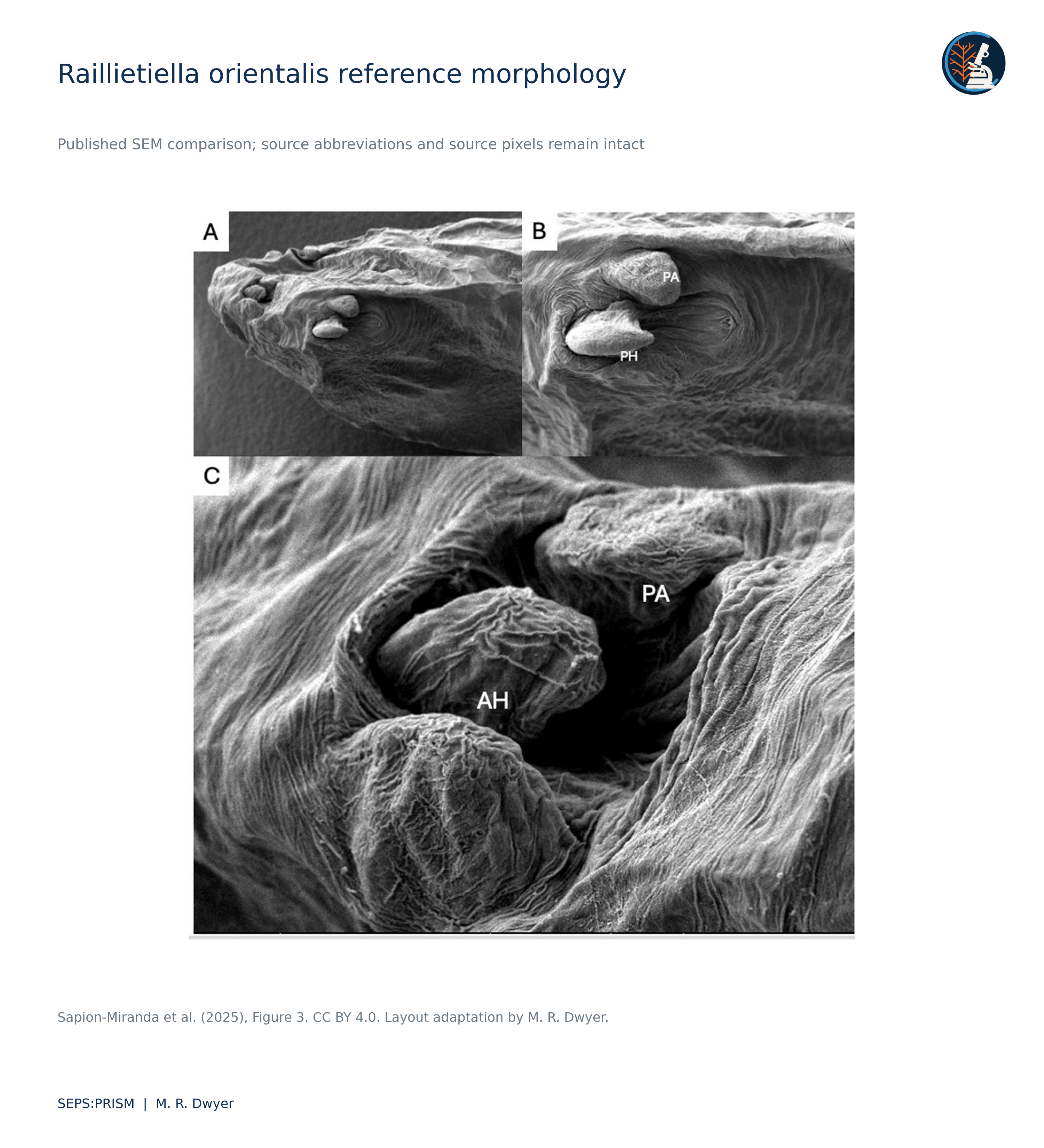
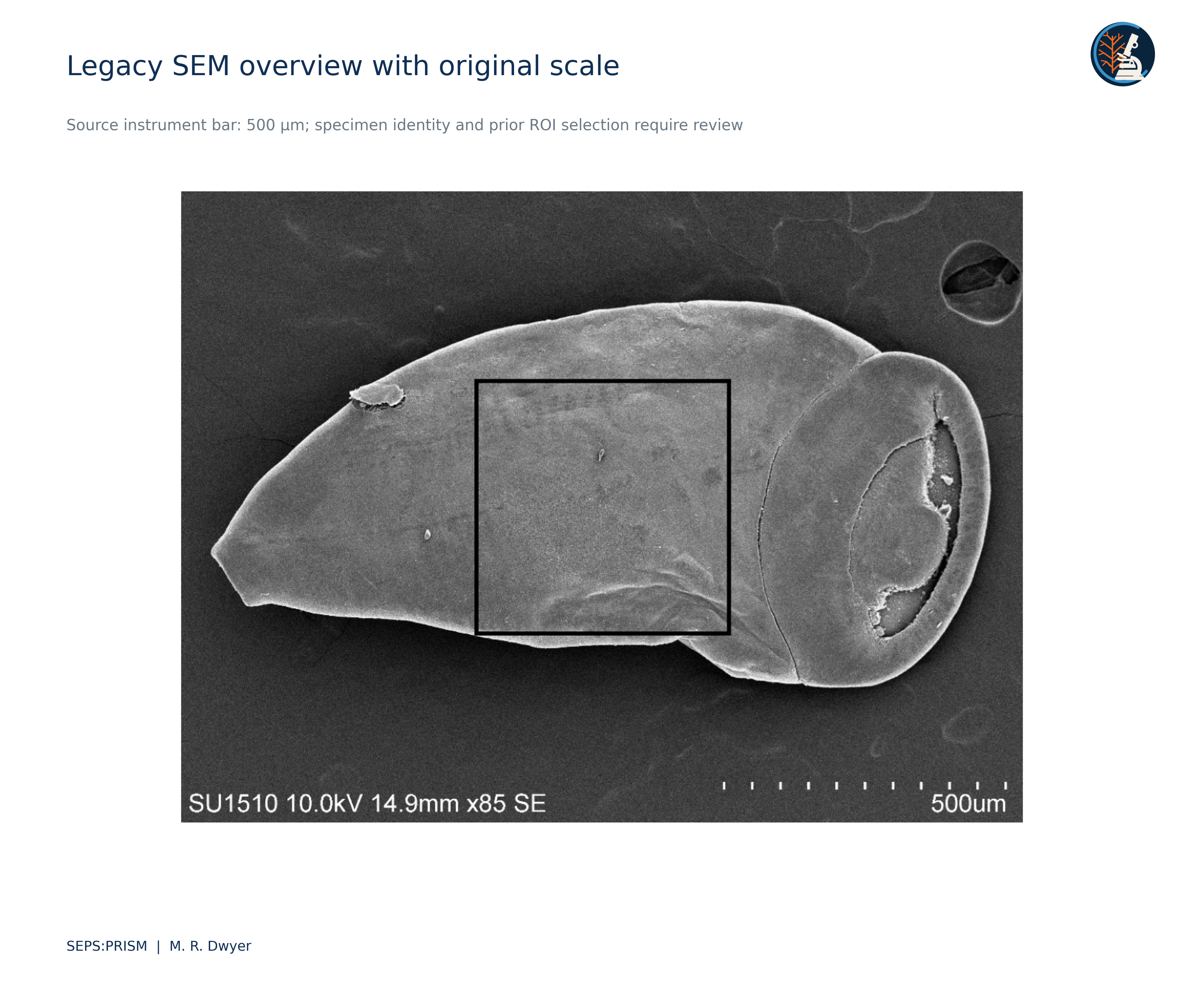
We reconcile two visually similar but biologically distinct scanning electron microscopy sources. The multipanel montage depicts Linguatula serrata from Shamsi and colleagues, whereas the three-panel image depicts Raillietiella orientalis from Sapion-Miranda and colleagues. We retain their identities and source attributions. A recorded endpoint pair and a 100 micrometer scale bar in the Linguatula montage yield a projected separation of 137.18 micrometers. A selected endpoint and bar perturbation produces a sensitivity range of 131.02 to 143.52 micrometers, which we do not interpret as a confidence interval. The source comparison supports careful character documentation and illustrates why a measurement can be reproducible while its anatomical interpretation still requires review. We also identify conflicting scale overlays in three legacy SEM figures, retire the unsupported dimensions, and recover an overview that retains its original 500 micrometer instrument bar.
The recovered montage belongs to a published Linguatula serrata study (Shamsi et al., 2020). The separate three-panel image belongs to a study of Raillietiella orientalis in a captive Meller’s chameleon (Sapion-Miranda et al., 2025). We therefore keep the two sources in separate records. Neither becomes an image of an unpublished SEPS specimen because it appeared in a SEPS presentation.
The three-panel reference identifies anterior and posterior hooks and a cephalic papilla using the source abbreviations AH, PH and PA. We preserve that attribution and avoid adding unsupported species-diagnostic labels. Occlusion and unavailable local calibration prevent a complete hook measurement from the recovered display. We use the source image for comparative orientation rather than create a full-hook dimension from a visible partial structure.
The retained measurement record selects endpoints (1030, 863) and (1220, 835) in the original montage coordinate system. Their separation equals √(190² + 28²) = 192.0521 pixels. The source scale bar extends from (1222.5, 1133) to (1362.5, 1133), a length of 140 pixels labeled 100 micrometers. We retain these coordinates and the original source image. An anatomical reviewer must confirm that the endpoints represent the intended pore-related structures before the distance acquires a specific biological label.
 (1)
 (2)
For a transparent sensitivity calculation, we allow each endpoint to move within a radius of 3 pixels and each scale-bar end within 1 pixel. The pair distance can then change by at most 6 pixels, and the bar length by at most 2 pixels. Applying the extreme numerator and denominator combinations yields the following bounds.
 (3)
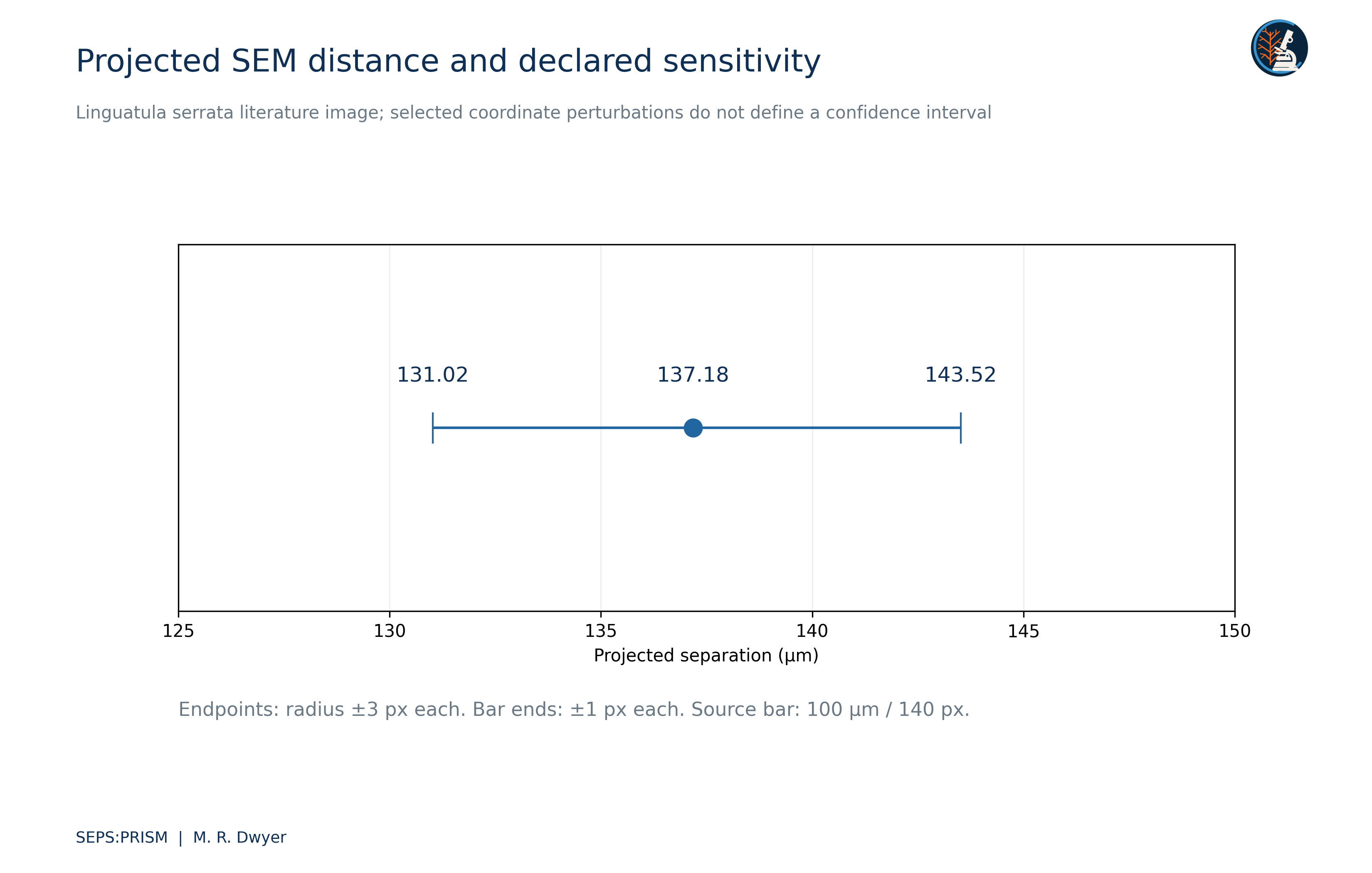
These bounds express selected geometric perturbations. They do not estimate observer error, instrument calibration error, specimen shrinkage or three-dimensional surface distance. The SEM projection and specimen preparation can influence the relationship between the measured image separation and the biological structure. We therefore report projected separation and preserve the scenario definition.
We would record the view, developmental state, sex when known, preparation and source scale for each diagnostic structure. A hook tip without its base supports a different set of measurements from a complete hook. A surface feature that appears in one view may disappear through orientation or coating. SEPS:PRISM therefore links the character decision to the exact region rather than to a general resemblance between montages (Dwyer, 2026a).
The corrected sources allow a useful reference comparison without merging taxa or pretending that display quality establishes identification. We preserve the literature figure as comparative evidence, the numerical record as a repeatable image-space calculation and the anatomical determination as a separate review task. This separation also allows students to inspect why a clearly rendered measurement may still have a limited biological interpretation.
The Shamsi source carries a Creative Commons Attribution NonCommercial NoDerivatives license. We retain the source for research review and exclude a modified image plate from the public release set pending appropriate permission. The report can communicate the calculation and citation without republishing an altered montage. The Sapion-Miranda source carries a Creative Commons Attribution license; the accompanying reference plate retains source credit and identifies the layout adaptation. We keep all source images distinct from SEPS-acquired specimens.
We inspected the supplied funding-pitch derivatives and the embedded images in the associated integrated proposal (Dwyer, 2025). The overview retains a 500 micrometer instrument scale but adds a 200 micrometer label over its lower-right corner. The texture view retains a 50.0 micrometer instrument scale and adds a 1 micrometer label. Neither added annotation provides a traceable pixel-length calculation that supports the displayed claim. Different scale lengths can coexist legitimately, but each requires its own correctly proportioned bar. We therefore retire the added labels and retain the original acquisition scales.
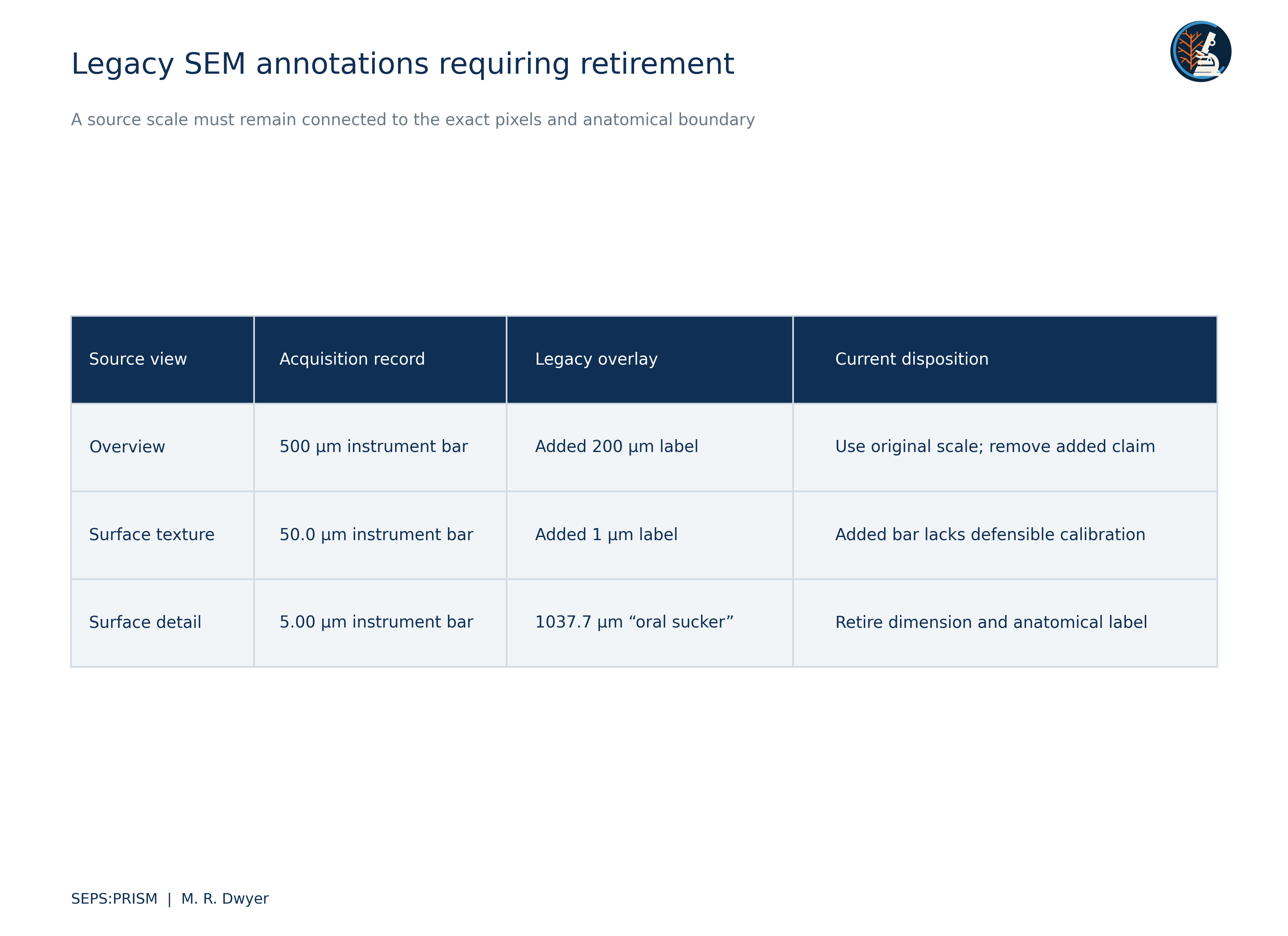
The file labeled oral sucker shows a high-magnification granular surface, a 5.00 micrometer instrument scale, and a full-frame line labeled 1037.7 micrometers. The selected line does not delimit a documented oral-sucker boundary, and its numerical value conflicts with the displayed source scale. We withdraw both the dimension and the anatomical assignment. This image may still preserve useful surface detail, but it cannot support the previous oral-sucker measurement.
 (4)
A valid replacement requires a source-bar length b measured in the same raster coordinate system as the feature distance d. Proportional resizing preserves this ratio; an undocumented crop, stretch or reused calibration can break it. We recovered a less altered overview from the integrated proposal and preserve its 500 micrometer instrument scale and existing black region box. We assign no replacement oral-sucker dimension or taxonomic identity. The source record must establish the specimen and acquisition context before a specialist interprets the anatomy.
Dwyer, M. R. (2025). UF integrated parasite identification proposal and funding-pitch image derivatives [Unpublished project materials; supplied archive SRC069 and SRC071]. Society for Evolutionary Parasite Systematics.
Dwyer, M. R. (2026a). SEPS:PRISM methodology reference (Version 1.2, 18 October 2026) [Methodology specification]. Society for Evolutionary Parasite Systematics.
Sapion-Miranda, P., Ebmer, D., Kniha, E., Walochnik, J., Dreyer, S., Fischer, D., Grund, L., Taubert, A., Hermosilla, C., & Hallinger, M. J. (2025). First identification of a patent pentastomid pulmonary (Raillietiella orientalis) infection in a captive Meller’s chameleon (Trioceros melleri) in Germany. International Journal for Parasitology: Parasites and Wildlife, 26, 101045. https://doi.org/10.1016/j.ijppaw.2025.101045
Shamsi, S., Barton, D. P., Zhu, X., & Jenkins, D. J. (2020). Characterisation of the tongue worm, Linguatula serrata (Pentastomida: Linguatulidae), in Australia. International Journal for Parasitology: Parasites and Wildlife, 11, 149–157. https://doi.org/10.1016/j.ijppaw.2020.01.010