 SEPS
Society for Evolutionary Parasite Systematics
SEPSM. R. Dwyer
Research and technical report. No validated end-to-end identification performance claimed.
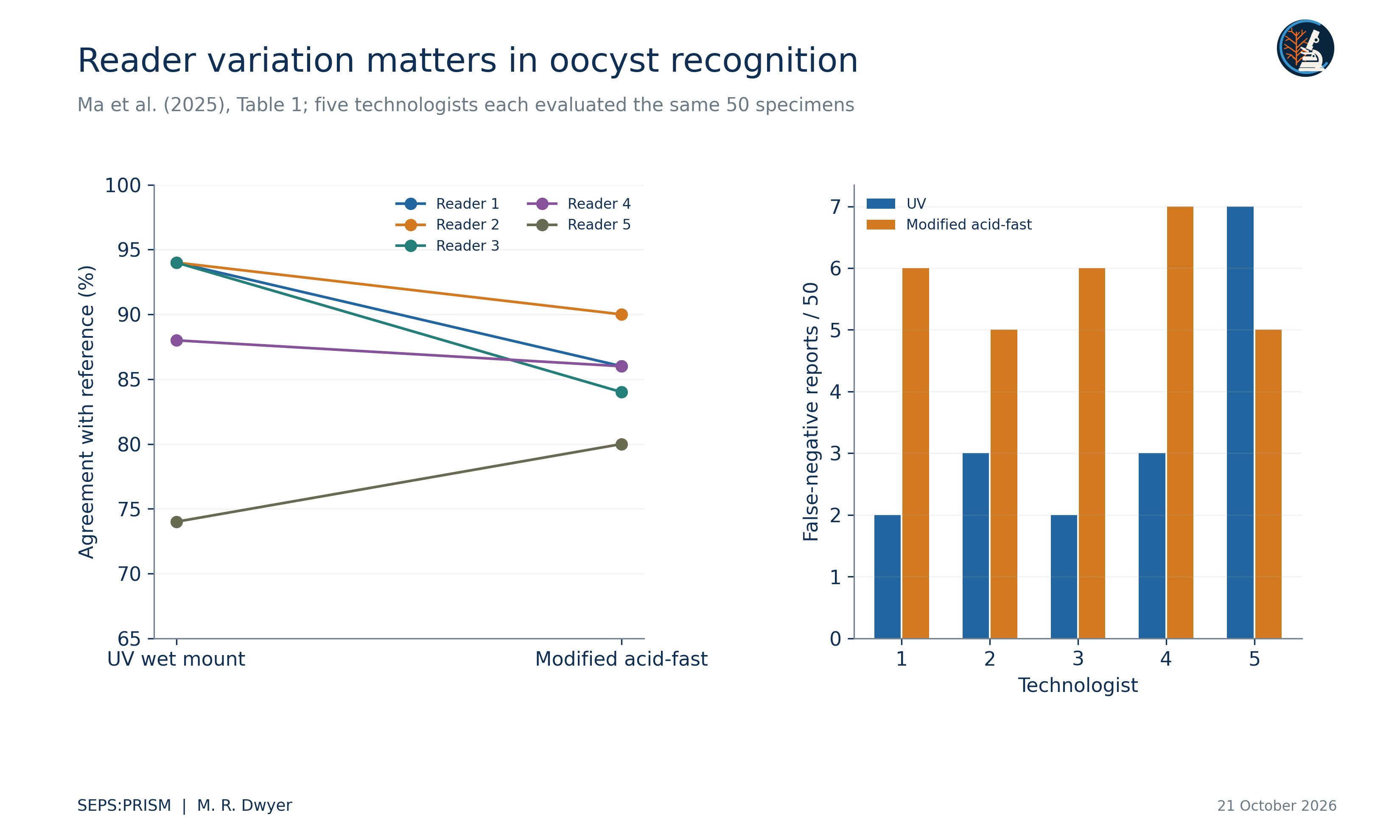
We examine how a microscopy reference can support recognition without implying viability, infectivity or validated automated diagnosis. We reconstruct the five-reader comparison reported by Ma and colleagues and pair it with authenticated CDC DPDx images. Across five readers, ultraviolet fluorescence agreement ranges from 74% to 94%, whereas modified acid-fast agreement ranges from 80% to 90% on the same 50-specimen set. Reader dependence, shared specimens and differences among biological endpoints prevent a single accuracy value from summarizing the problem. We specify source-linked image records and a prospective paired evaluation within SEPS:PRISM. The resulting gallery and reader comparison support education and study design; they do not establish clinical performance of SEPS.
A microscopy image records appearance under a particular preparation and optical configuration. We interpret a visible wall, internal bodies or fluorescence through that configuration and the relevant comparative material. We distinguish recognition of an oocyst-like structure, taxonomic identification, developmental state, viability and infectivity. Evidence for one endpoint does not automatically establish another. The CDC DPDx reference provides comparative morphology and preparation context for Cyclospora cayetanensis (Centers for Disease Control and Prevention [CDC], 2019).
We revised the supplied atlas around this distinction. An unrelated whole-worm image cannot illustrate an oocyst trait, and a mixed-taxon image requires labels that preserve both organisms. We therefore replace the legacy gallery with six source-authenticated CDC panels. We keep their source arrows and identify Cryptosporidium parvum separately in the safranin comparison. The panels show different specimens and methods rather than a time series or repeated imaging of one oocyst.
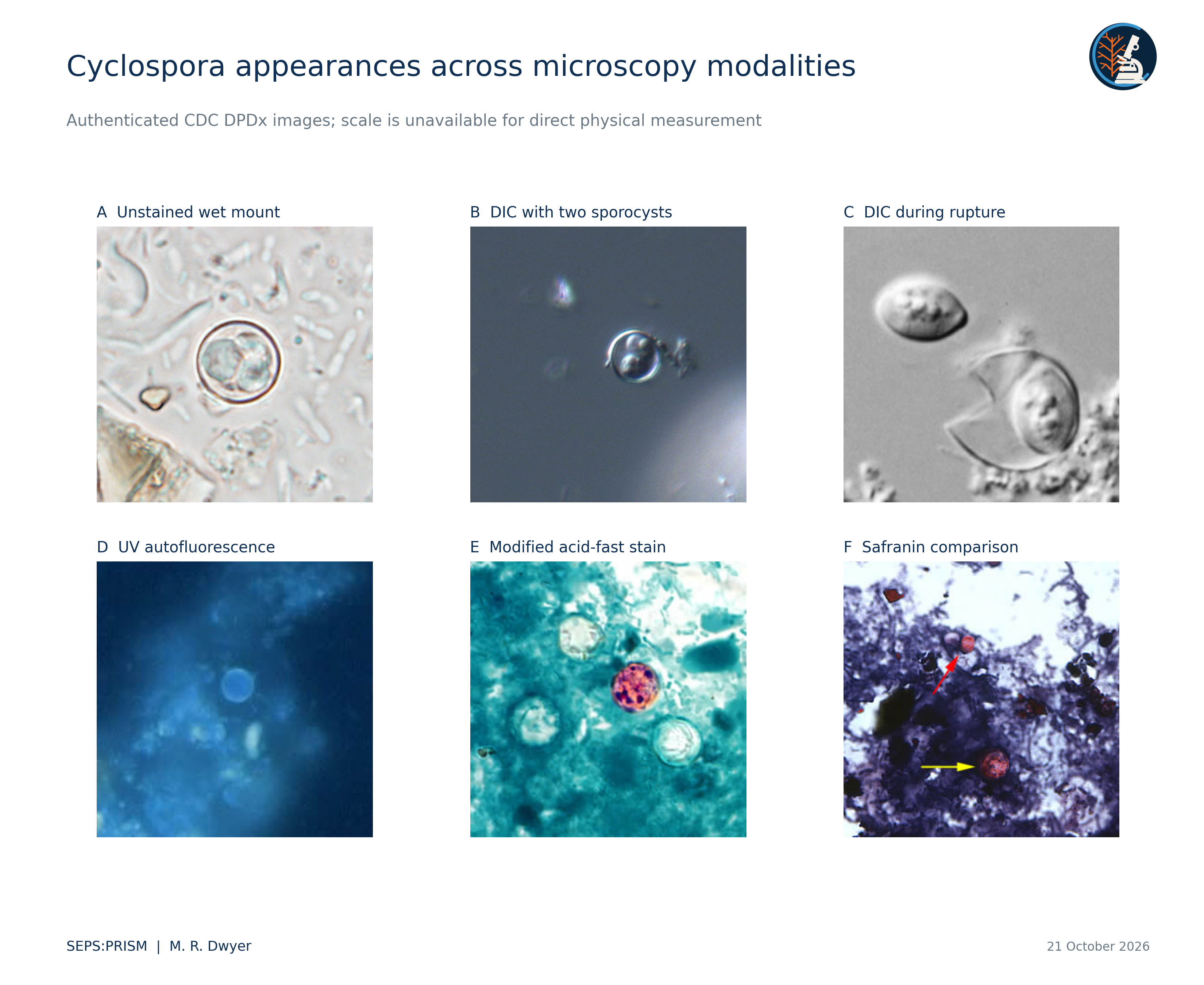
We retain each source file, original filename, preparation label and source URL. We do not estimate a physical dimension from the displayed diameter of a downloaded image. A microscope magnification label cannot resolve later resizing. A measured pixel diameter can describe that file, but a physical size comparison requires a verified scale in the same coordinate system. We also avoid estimating stain concentration or fluorescence intensity from differently exposed reference images.
For future acquisition, we would record objective, numerical aperture, illumination or excitation configuration, camera settings, preparation and calibration image alongside the specimen identifier. These fields distinguish biological variation from acquisition variation. We would preserve unprocessed frames and describe any background correction mathematically. We would never remove a weakly stained object merely to improve the visual consistency of a training set.
Ma et al. (2025) compared five technologists on the same 50 specimens: 35 Cyclospora-positive, five Cystoisospora-positive and ten negative specimens. We transcribe their table rather than digitize a chart. Ultraviolet agreement counts are 47, 47, 47, 44 and 37; modified acid-fast counts are 43, 45, 42, 43 and 40. The method differences vary by reader. These repeated evaluations retain 50 independent specimen identities, not 250 independent specimens. The figure reports false-negative counts per reader, without relabeling the source denominator as the number of infected specimens.
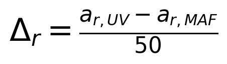 (1)
The paired agreement differences for readers 1 through 5 equal 0.08, 0.04, 0.10, 0.02 and −0.06. These are descriptive reader-specific contrasts. The aggregate table does not reveal which individual specimens produced discordant outcomes, so it cannot supply the paired contingency counts for a specimen-level McNemar calculation.
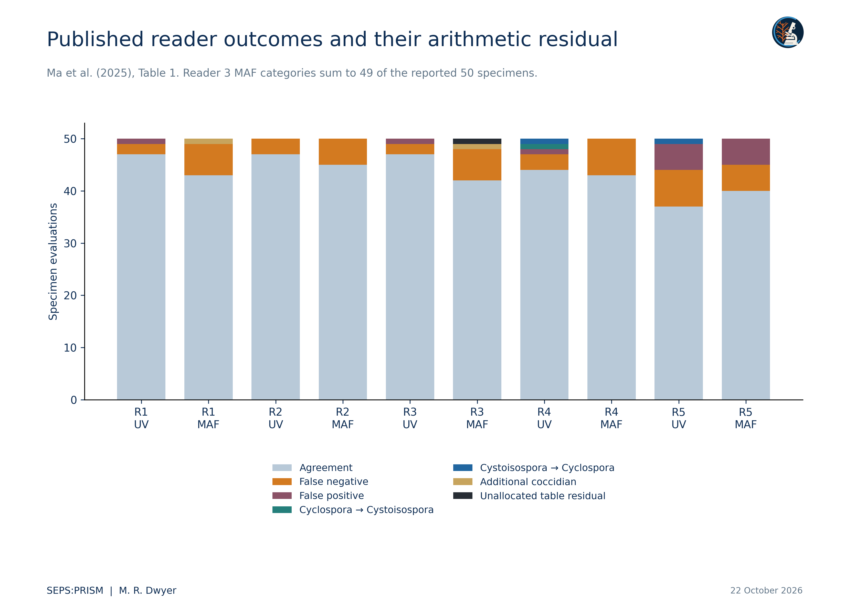
The source table also distinguishes incorrect coccidian assignments and reports of an additional coccidian. Those categories account for the observations that agreement, false negatives and false positives alone do not explain. For ultraviolet microscopy, reader 4 has two taxon-discrepant results and reader 5 has one. For modified acid-fast microscopy, readers 1 and 3 each report one additional coccidian. Nine reader-method totals then equal 50. Reader 3 under modified acid-fast microscopy still sums to 49: 42 agreement, six false negatives and one additional-coccidian report. The source leaves an arithmetic residual of one evaluation. We display that residual without assigning an outcome or silently correcting the published count. We preserve the source category definitions instead of forcing a binary confusion matrix (Ma et al., 2025).
 (2)
Here Y_irm indicates correct classification of specimen i by reader r under method m. A specimen effect u_i represents shared difficulty, a reader effect v_r represents reader variation, and w_rm permits reader-specific method response. This equation specifies a possible crossed design. We have not fitted it to the aggregate table and make no claim about its coefficients.
A prospective study should randomize reading order, preserve washout where appropriate and retain every individual decision. It should define the adjudicated reference before analysts inspect model outputs. The unit of partitioning should keep all images of a specimen together, and the external evaluation should include different acquisition conditions. We would report class-specific errors and unresolved decisions alongside agreement, with uncertainty that respects the repeated-reader design.
 (3)
This identity shows why a sensitivity and specificity pair does not determine positive predictive value without a target prevalence π. We include no fitted numerical example because the recovered image archive supplies neither a population prevalence nor SEPS sensitivity and specificity estimates.
FieldView or laboratory import captures source context. TEGUMENT retains object boundaries and difficult background regions. HookFuse computes only features whose optical basis we can justify. FUZZARIA records missing calibration, preparation mismatch and disagreement. Expert review determines the final reporting level (Dwyer, 2026a). We retain failed and unresolved cases when estimating system coverage. A useful teaching example can remain unresolved at species level while clearly demonstrating why a particular image is insufficient.
Centers for Disease Control and Prevention. (2019, April 10). DPDx cyclosporiasis. https://www.cdc.gov/dpdx/cyclosporiasis/index.html
Dwyer, M. R. (2026a). SEPS:PRISM methodology reference (Version 1.2, 18 October 2026) [Methodology specification]. Society for Evolutionary Parasite Systematics.
Ma, A., Mathison, B. A., & Couturier, M. R. (2025). Shedding new light on Cyclospora: How the use of ultraviolet fluorescence microscopy can improve diagnosis of cyclosporiasis. Journal of Clinical Microbiology, 63(1), e01084-24. https://doi.org/10.1128/jcm.01084-24