Clean, carefully composed imagery for exploration and education.
Morphology · Collections · Imaging · Systematics
Society for
Evolutionary
Parasite Systematics
Advancing parasite systematics through field biology, specimen-linked imaging, computational tools, and collaborative research.
THE SPECIMEN COMES FIRST
Anatomy is the starting point.
Detailed microscopy makes parasite structures visible and gives comparative research something concrete to examine.
Explore how we document morphology →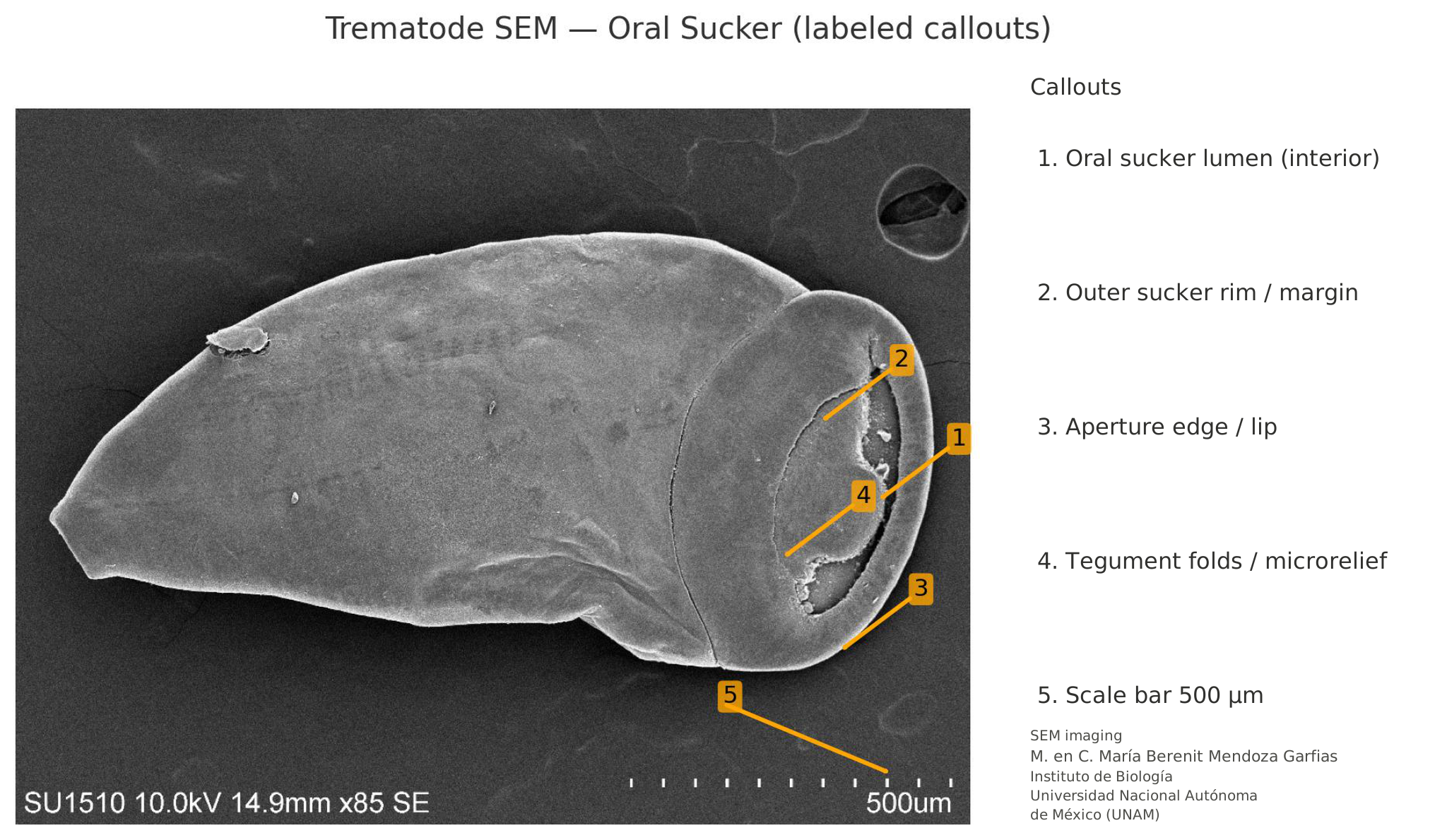
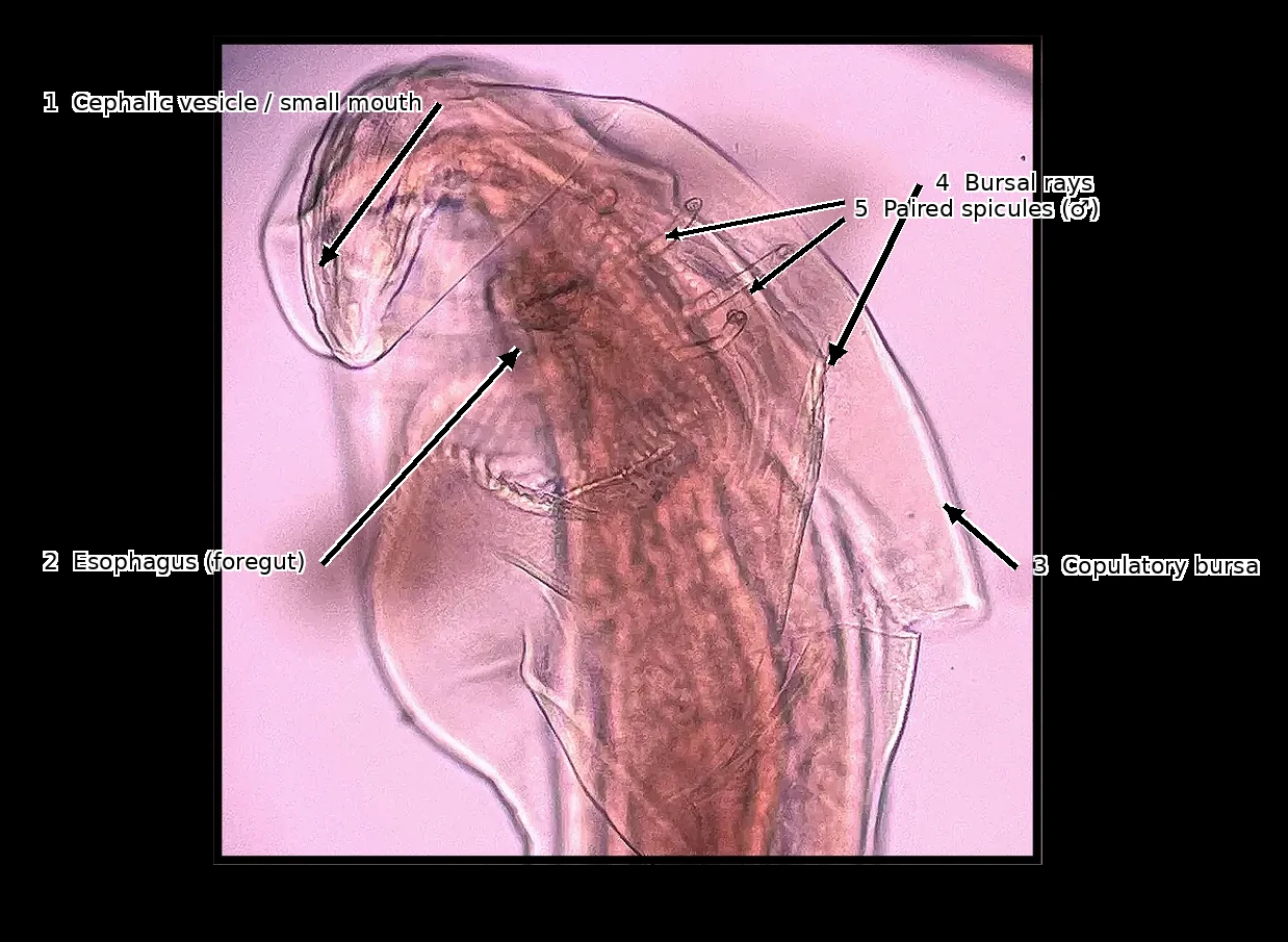
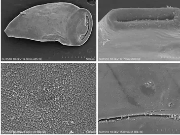
Figure 1 · Trematode under SEMSupplied anatomical annotations shown for illustration; the original instrument labels are retained.SEM imaging: M. en C. María Berenit Mendoza Garfias · Instituto de Biología, Universidad Nacional Autónoma de México (UNAM).
Open full figure ↗Mission:
To understand parasite diversity and its place in changing host communities.
SEPS is a nonprofit research organization focused on parasite systematics and disease ecology. Our work connects field observations with comparative morphology, taxonomy, and preserved specimens to investigate neglected and understudied parasites. Computer vision supports this research by organizing images and measurements for expert review.
Building on this foundation, we aim to advance parasite systematics and translate that knowledge into affordable, accessible imaging tools that strengthen diagnostic capacity. Our long-term goal is to develop and validate approaches suited to settings with limited laboratory infrastructure, including those equipped with a smartphone and a microscope, to support reliable parasite identification and efforts to combat parasitic neglected tropical diseases worldwide.
EXPLORE
From specimen to record.
The images, diagnostic structures, and documentation behind our research.
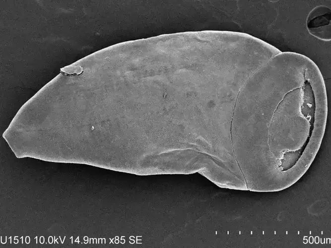 Specimen archiveWhole body → detail
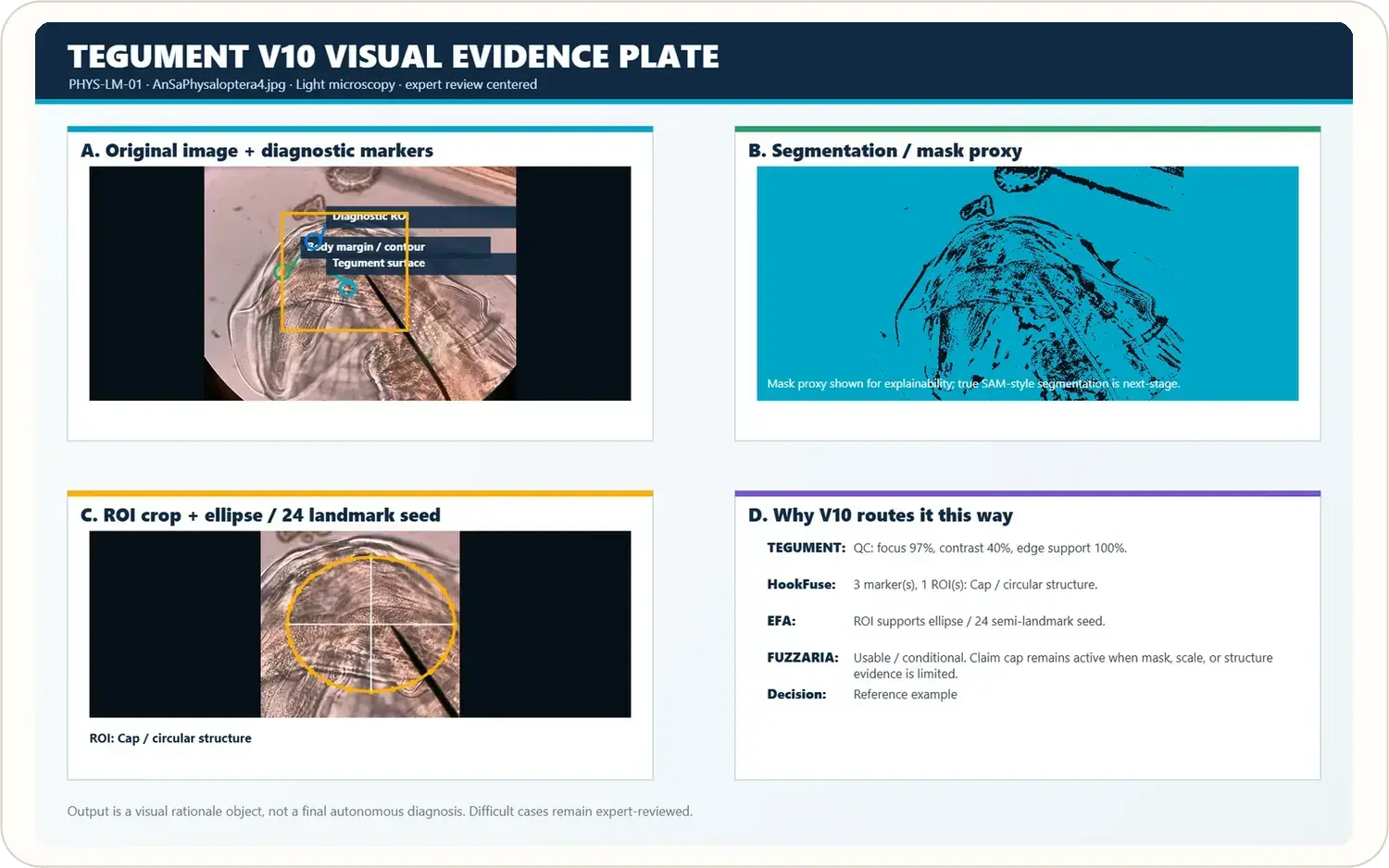 Evidence plateSee the rationale
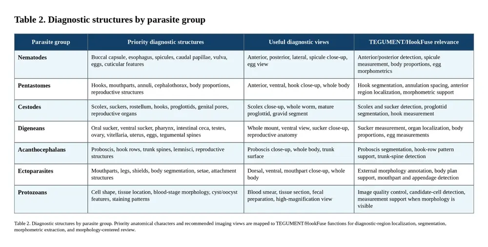 Diagnostic structuresViews + characters
 Morpho-BarcodeOne linked record
SEM imaging: M. en C. María Berenit Mendoza Garfias · Instituto de Biología, UNAM.
THE STANDARDEvidence first.
Evidence first.
Confidence earned.
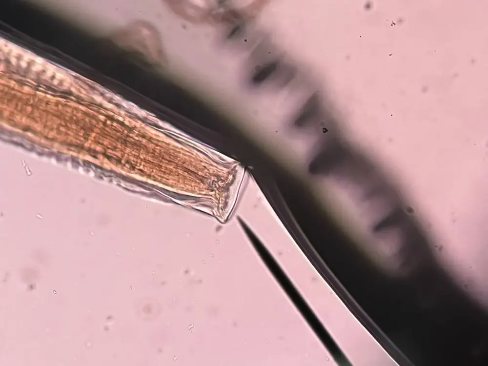
One nematode photograph connects the original capture, proposed labels, and human review.
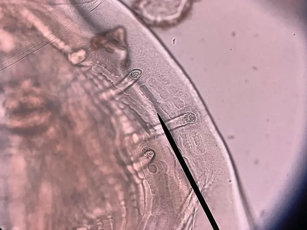 01 ORIGINAL CAPTURERaw stays rawPreserve the source. Keep masks, outlines, and other derivatives linked to it.View original capture ↗
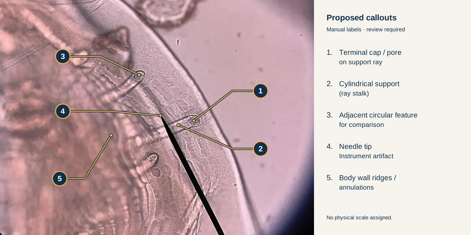 02 EVIDENCE & LIMITSClaims stay cappedLabel visible structures, keep uncertain assignments provisional, and state the limits.View annotated plate ↗
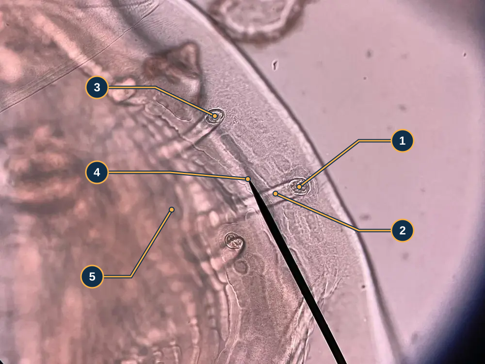 03 EXPERT INTERPRETATIONReview stays humanOpen a number, compare the proposed label, and record your review notes.Open numbered review ↗
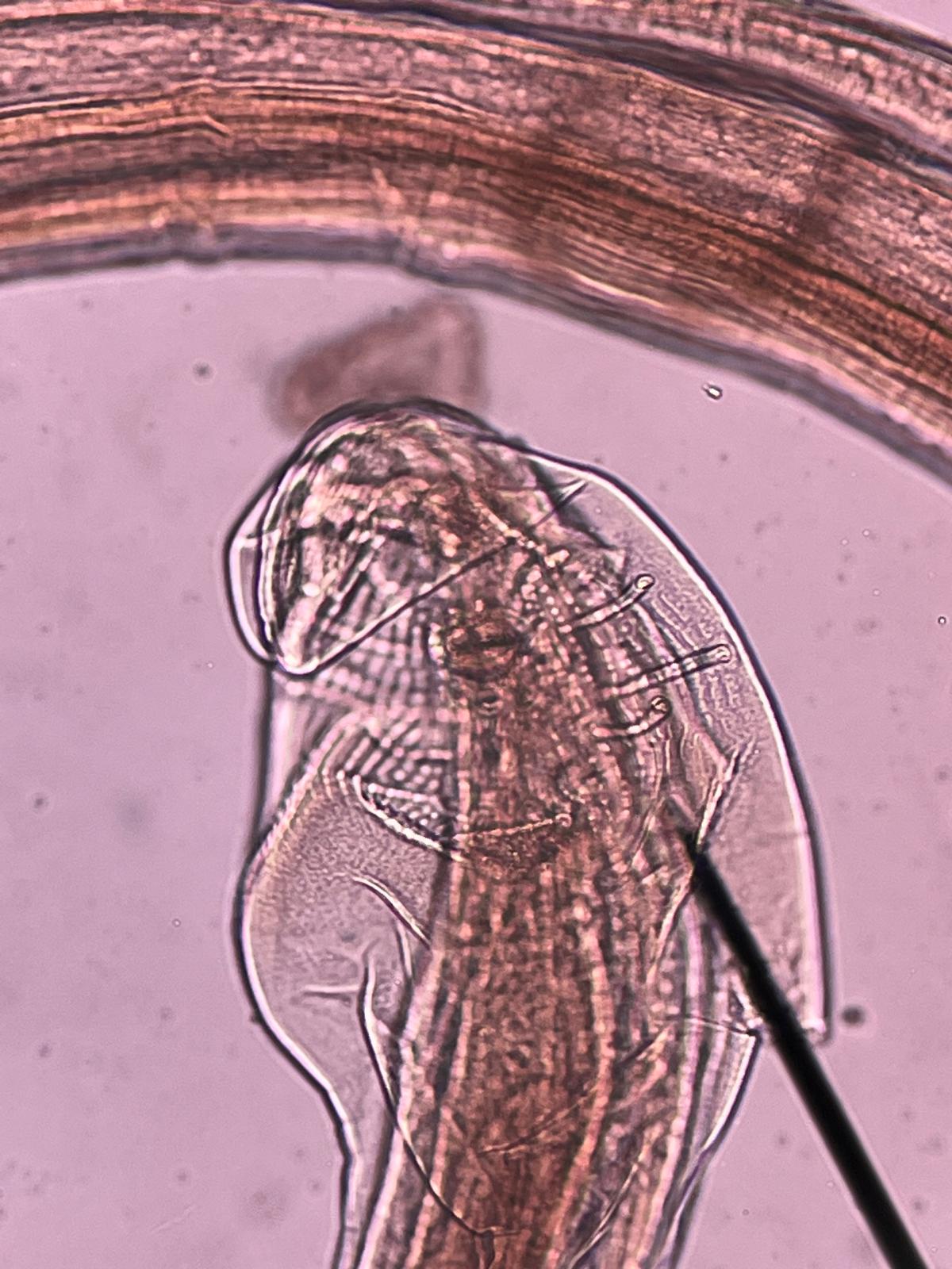
All three views use the same archive photograph. Labels remain provisional; the needle is an artifact, and no physical scale or species identification is assigned.
OPEN RESEARCH COLLECTION
Open research collection Explore the evidence behind the work.
Technical reports and proposed laboratory pilots connect parasite systematics with inspectable evidence. Explore documented analyses alongside new research concepts for freshwater snail trematodes and mealworm gregarines.
Inaugural specimen
The organism comes first.
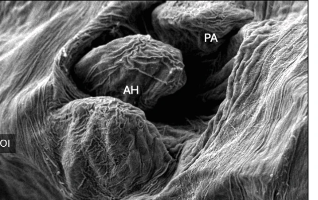
This anterior view opens the SEPS digital collection—and captures the central purpose of SEPS: making parasite morphology visible, interpretable, and enduring.
Public specimen pages will pair gallery-quality imagery with scientific versions retaining scale, annotation, preparation details, host context, evidence status, and voucher linkage whenever available.
Read the SEPS:PRISM methodology ↗
Research program
Field evidence, specimens, and evolutionary questions.
SEPS studies parasite diversity and movement across hosts and landscapes, with particular attention to invasive systems, emerging risks, and the diagnostic morphology needed to recognize them.
The digital collection
A visual reference built to last.
Scale, labels, annotations, metadata, and review state retained.
What supports an interpretation—and what still limits it.
Research instruments
Software serves the specimen—not the other way around.
SEPS:PRISM means Parasite Recognition through Integrated Specimen Morphology. Its components organize images, review diagnostic structures, preserve uncertainty, and support expert interpretation.
TEGUMENT
The first processing phase within SEPS:PRISM: image preparation, specimen segmentation, and boundary review.
HookFuse
Brings selected structures, life stage, orientation, and specimen context into one review surface.
FUZZARIA
Weights evidence, preserves limitations, and helps prioritize expert review.
FieldView
Mobile-first capture for ectoparasite observations, host context, raw field imagery, and reviewable evidence.
Swipe or drag sideways4 modules
Explore the SEPS:PRISM workflow ↗
Help build the reference collection parasites deserve.
SEPS welcomes collaboration with taxonomists, museums, field biologists, microscopists, veterinarians, molecular laboratories, agencies, educators, and students.
Research program
Parasite systematics across organisms, images, and ecosystems.
Documenting how parasite form, host association, life stage, and movement become quantitative, reviewable evidence.
Parasite biodiversity discovery
Diagnostic morphology, high-resolution imaging, specimen metadata, and voucher linkage support evaluation of poorly characterized morphotypes, locality records, host associations, and candidate diversity.
Invasive species & parasite movement
SEPS distinguishes co-introduction, enemy release, native-parasite acquisition, host switching, spillover, and plausible spillback pathways rather than treating every novel association as the same process.
Host–parasite ecology & coinfection
Host identity, size, sex, condition, habitat, season, infection site, richness, intensity, and coinfection structure remain connected to the underlying specimen records.
Quantitative morphology
Landmarks, outlines, hook geometry, annulation, body proportions, attachment structures, and surface texture are compared only when orientation, scale, and processing history are controlled.
Progressing morphology
From descriptive characters to trajectories.
A single striking specimen is not a developmental series. Replication, standardized views, calibration, and repeatability remain the gatekeepers.
Length-to-width relationships, outline, regional proportions, annulation, and curvature.
Hook or sucker geometry, spacing, curvature, insertion angle, and asymmetry.
Surface texture and microstructure add evidence but never rescue an inadequate diagnostic view.
Diagnostic imaging
Acquire the structure before measuring it.
Useful images are not automatically diagnostic. Orientation, completeness, scale, focal plane, and visible character support determine what a record can responsibly say.
Parasite burden is more informative when the host is measured too.
SEPS evaluates condition metrics alongside body size, sex, season, site, reproductive state, and species-specific allometry. Association is reported before mechanism is implied.
PrevalenceProportion of hosts infectedIntensityParasites per infected hostConditionValidated host-state estimate
Active and emerging projects
Questions large enough to matter. Claims narrow enough to defend.
SEPS organizes projects around biological questions, specimen evidence, and explicit validation gates.
Archive active
Analysis in progress
Validation required
Long-term program
Pentastome comparison and host movement
Comparative records integrate host identity, anatomical site, life stage, prevalence, intensity, richness, coinfection, morphology, voucher status, and confirmation evidence.
Core question
Which associations represent co-introduction, spillover, native-parasite acquisition, host switching, or a plausible spillback pathway?
Cyclospora: oocyst walls and detection
Research on the Cyclospora cayetanensis oocyst wall, asking how its structure relates to environmental transmission, diagnostic appearance, and the challenges of detection and control. Direct evidence is distinguished from inference based on other coccidia.
Core question
How can standardized imaging, explicit uncertainty, and clear confirmation pathways improve oocyst recognition and surveillance readiness?
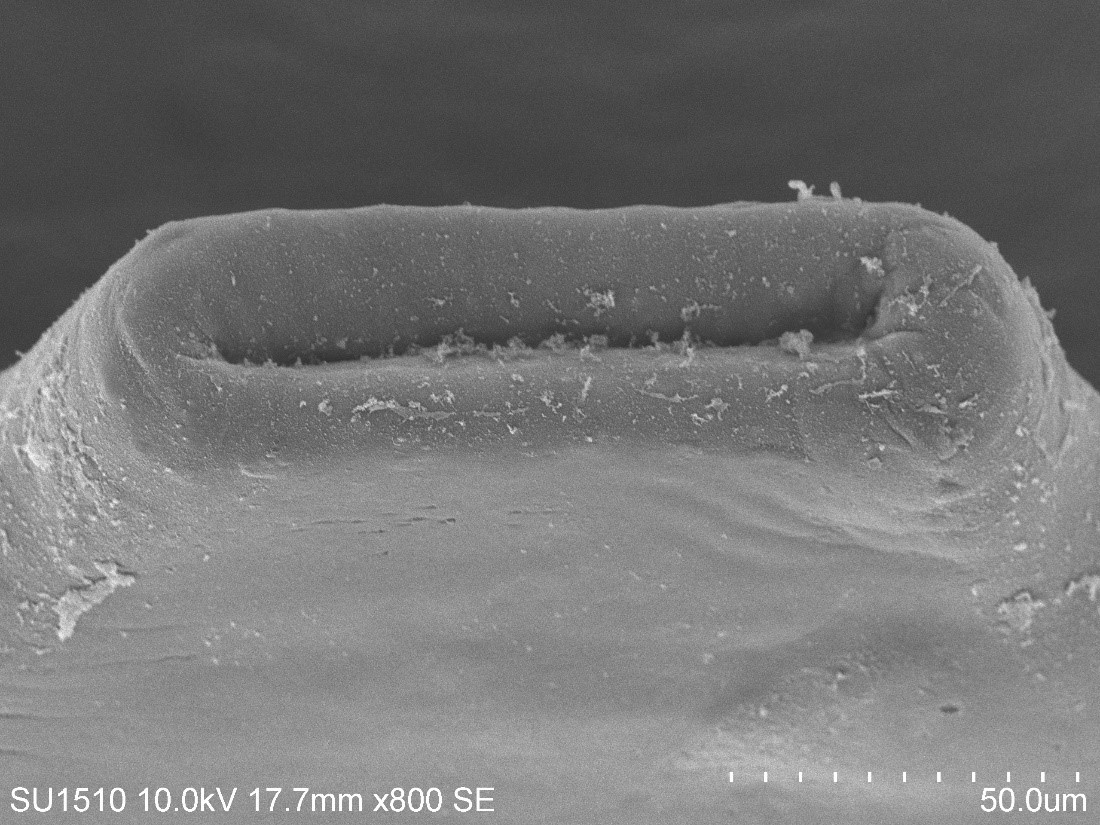
SEM imaging: María Berenit Mendoza Garfias, Instituto de Biología, Universidad Nacional Autónoma de México (UNAM).
Parasite morphology across life stages
Comparative imaging of parasite form across life stages, with attention to body organization, attachment structures, and surface features. The goal is to connect documented morphology with developmental context and features useful for identification.
Core question
Which changes reflect development or individual variation, and which features help distinguish parasite groups?
Recall-first microscope-field localization
A candidate-proposal system is evaluated by field-level recall, review burden, category capture, failure modes, and external challenge performance—not inflated diagnostic language.
Core question
Can heterogeneous fields be triaged reliably enough to make expert review faster without hiding misses?
New World screwworm: Biosecurity Without Panic
A research initiative on Cochliomyia hominivorax linking transmission, ecosystem effects, coinfection, and control. Proposed workflows use larval and adult morphology to strengthen visual evidence, conservative screening, and expert confirmation.
Core question
How can better specimen evidence support prompt official reporting and expert review while reducing confusion with look-alike flies?
South Florida specimen–image atlas
A durable regional archive linking hosts, collection events, parasite specimens, images, measurements, vouchers, sequences, review status, and publications.
Core question
Can scattered records become a surveillance network without losing taxonomic or specimen-level context?
Imaging & methods
Make morphology visible, measurable, and traceable.
The image is evidence only when acquisition, specimen, scale, processing, and interpretation stay connected.
Specimen first
Host, locality, infection site, preparation, life stage, voucher status, and physical specimen remain the center of the record.
Raw image preserved
Original files remain immutable while crops, masks, annotations, measurements, and enhanced derivatives are versioned.
Limits retained
Missing scale, incomplete views, damage, debris, focus limitations, and uncertainty remain visible rather than edited away.
SEPS:PRISM IN PRACTICE
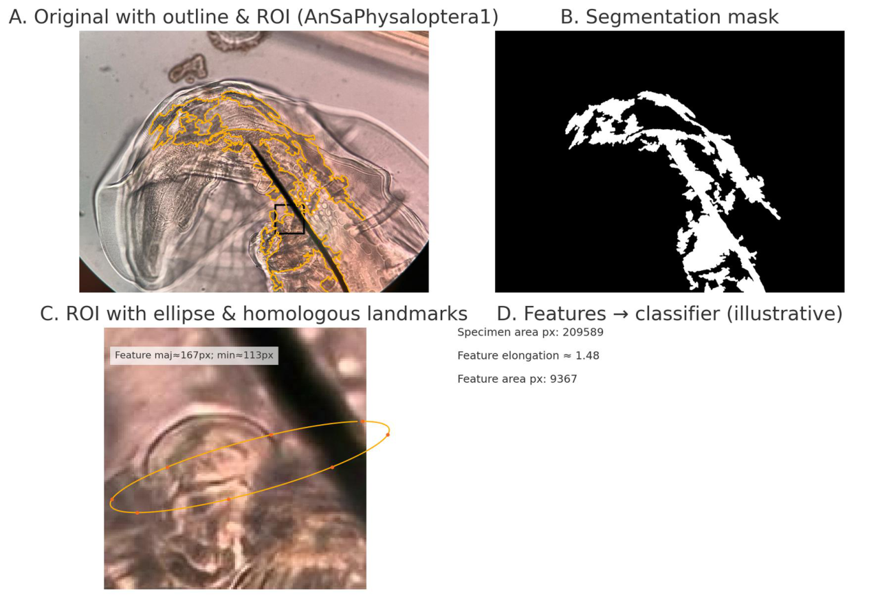
From image to outline.
Explore one specimen photograph, its provisional mask, and a mathematical description of the extracted boundary.
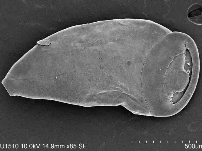
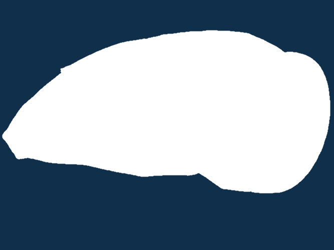
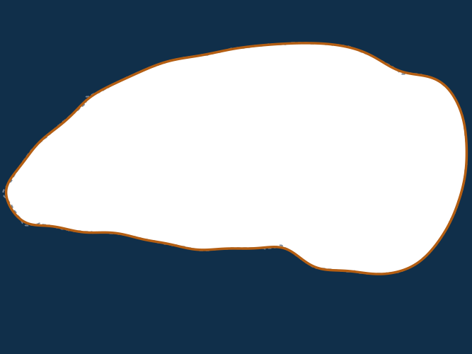
Extracted boundaryFourier approximation
From anatomical detail to a shape record.
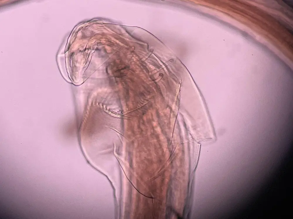
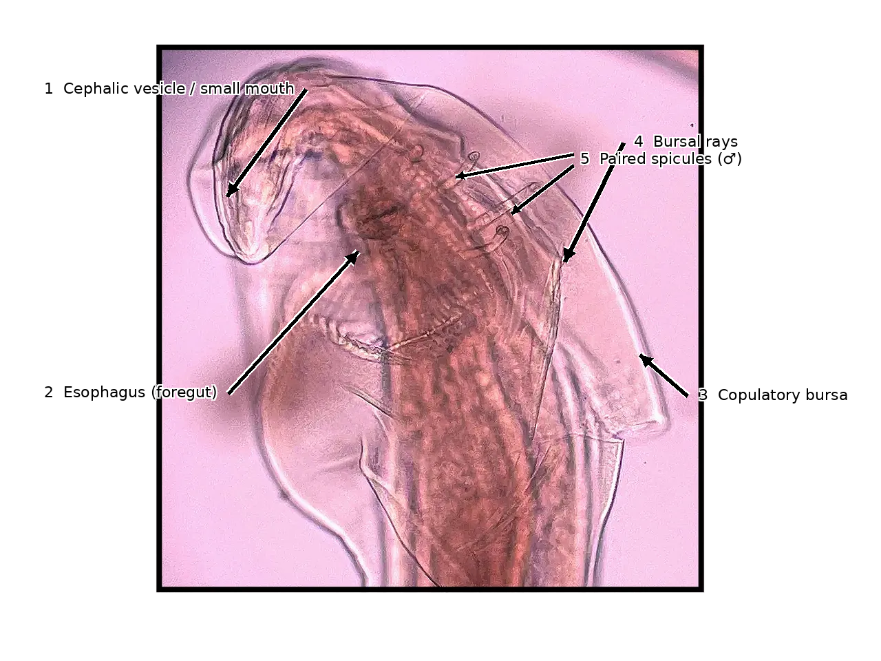
A close view documents the structures in an image. Outlines and landmark placements show different ways to record shape for comparison.
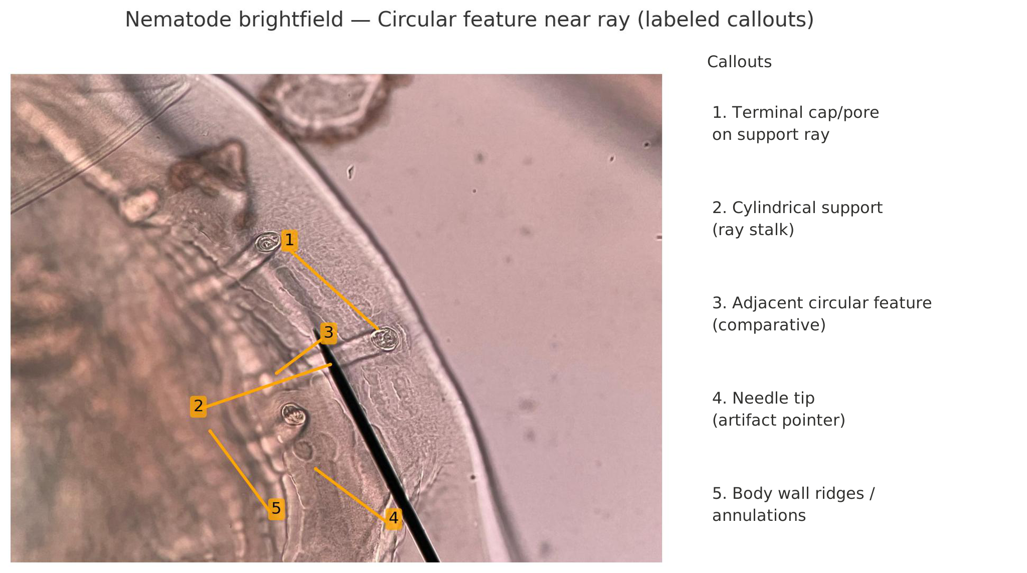
Figure 2 · A closer look at anatomyBrightfield microscopy with the supplied callouts spaced evenly for reading.
Open full figure ↗Illustrative examples. Anatomical labels and landmark correspondence require expert review.
Specimen-to-evidence path
One continuous chain.
- 01Field & host recordLocality, date, host context, condition, and collection event.
- 02Recovery & preservationAnatomical site, parasite count, life stage, preparation, and voucher pathway.
- 03Image acquisitionWhole body, diagnostic regions, scale, orientation, focal plane, and instrument metadata.
- 04Derived evidenceVersioned crops, masks, landmarks, measurements, annotations, and quality flags.
- 05Expert reviewInterpretation, limitations, comparison, and confirmation status.
Public collection modes
One specimen. More than one honest view.
See the organism.
The source photograph, preserved without annotation.
Read the record.
Proposed labels stay linked to the original. No calibrated scale is assigned.
Know the limits.
Compare the visible features and record what remains unresolved. Open the numbered review.
Operational companion
Test the image before the interpretation.
SEPS Lens checks focus signal, exposure, contrast, structure, segmentation readiness, and record integrity on the device in your hand.
ECOLOGICAL ANALYSIS WITHIN SEPS:PRISM
PARAPLEX
Parasite Assemblage and Relationship Analysis across Populations, Landscapes, and Ecological eXchange.
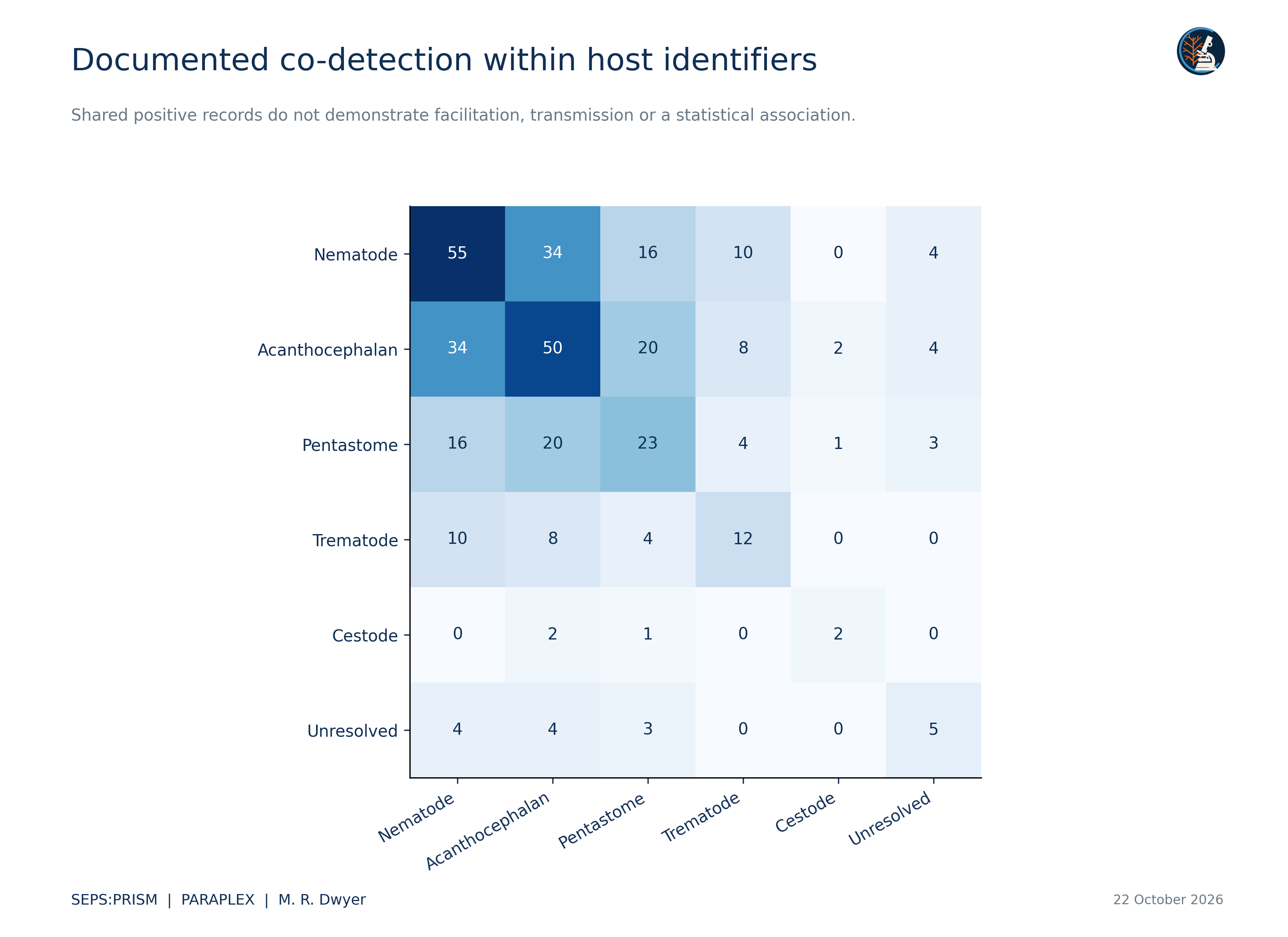
PARAPLEX connects reviewed specimen evidence to host and community questions. The current descriptive analysis preserves the original observation rows, summarizes recorded groups and reconciles candidate links. Population and transmission models require suitable examination records and sampling designs.
SEPS Lens · browser-native image evidence
See what the image can support.
See what the image can support.
Keep the limits visible.
Real computer-vision measurements and fuzzy evidence rules run locally. The result is an image-readiness assessment—not an organism identification.
On-deviceNo uploadNo autonomous ID
01
Acquire
Bring in the image.
Runs the entire analysis chain with a bundled real microscope image.
02
Inspect
Interrogate the pixels.
Measuring image evidence…
03
Reason
Fuzzy score. Explicit caps.
0/100
Awaiting image
No evidence assessment yet.
Load an image to calculate a transparent, non-diagnostic image-readiness score.
- Analysis pending.
- Analysis pending.
Open the decision trace +
Local history
Recent evidence checks.
Only scans you explicitly save appear here. They stay on this device until browser data is cleared.
No saved scans yet.Your first locally saved assessment will appear here.
The boundary matters
Image quality is not taxonomic certainty.
SEPS Lens evaluates whether pixels are suitable for morphological review. It does not replace keys, vouchers, scale, host and locality context, literature, molecular confirmation when appropriate, or expert interpretation.
The wider SEPS program
Built from fieldwork, necropsy, microscopy, and iteration.
An independent, morphology-first research organization focused on parasite diversity, host–parasite movement, specimen-linked evidence, and responsible computational support.
 ABOUT SEPS
SEPSParasite diversity.
Society for Evolutionary Parasite Systematics
U.S. 501(c)(3) nonprofit
Parasite diversity.
Documented carefully.
SEPS is a U.S. 501(c)(3) nonprofit research organization focused on parasite systematics and disease ecology. Comparative morphology, taxonomy, ecology, and preserved specimens guide our work on neglected and understudied parasites. Computer vision and quantitative shape analysis support these scientific questions.
Building on this foundation, we aim to advance parasite systematics and translate that knowledge into affordable, accessible imaging tools that strengthen diagnostic capacity. Our long-term goal is to develop and validate approaches suited to settings with limited laboratory infrastructure, including those equipped with a smartphone and a microscope, to support reliable parasite identification and efforts to combat parasitic neglected tropical diseases worldwide.
 Identification standardsWhat the evidence can support
Research programsSouth Florida flagship
 CollaborateSpecimens · images · expertise
 SEPS:PRISM methodologyFrom image capture to expert review
Integrated methodology
SEPS:PRISM
Parasite Recognition through Integrated Specimen Morphology. FieldView acquisition, TEGUMENT image preparation, HookFuse morphology, and FUZZARIA evidence assessment feed expert review.
Source images remain linked to reviewed masks, crops, and versioned derivatives.
Selected diagnostic structures are evaluated with life stage, scale, and orientation.
Evidence strength and reasons to withhold a claim travel together.

Keep SEPS with you
Install SEPS Mobile.
Add this companion to your home screen for a full-screen, offline-ready experience.
On iPhone or iPad: tap Share, then “Add to Home Screen.”