Society for Evolutionary
Parasite Systematics
Image becomes evidence.
We investigate parasite diversity and host associations through comparative morphology, preserved specimens, and reproducible visual evidence.
SEPS is a nonprofit research organization focused on parasite systematics and disease ecology. Our work connects field observations with comparative morphology, taxonomy, and preserved specimens to investigate neglected and understudied parasites. Computer vision supports this research by organizing images and measurements for expert review.
Florida nonprofit organization · U.S. 501(c)(3)
Which parasites occur in sampled hosts, and which anatomical characters support their identification?
How do parasite records vary among hosts and collection sites? Observed associations provide a starting point for testing transmission and host-switching hypotheses.
Do conventional measurements, geometric morphometrics, and computer vision add reliable identification value when compared with expert morphological review?
Explore the images, diagnostic structures, and documentation behind our research.
Field-to-voucher research infrastructure
Comparative morphology and curated visual evidence
Reproducible analysis through TEGUMENT, HookFuse and FUZZARIA
Measurable outputs
Preserved vouchers Traceable datasets Diagnostic image atlases Peer-reviewed records GIS/data products Agency-ready implementation tools
Field → image → evidence.
Field validation, voucher generation, reproducible data products and agency-ready outputs.
Morphology, measurement, and computer vision.
SEPS is developing a linked workflow in which comparative morphology, conventional measurements, geometric morphometrics, and computer vision contribute distinct evidence. Taxonomic interpretation remains grounded in diagnostic anatomy, reference literature, and expert review.
Each record connects the available images to the host, collection event, preservation history, and voucher. Missing information remains visible. Molecular evidence can be linked when it is available and appropriate to the question.
This page describes the integrated research framework. Vision Transformer and geometric morphometric extensions are proposed for evaluation. It does not report validated automated parasite identification or measured performance.
Record identity, recovery site, collection context, image provenance, and known limitations.
Preserve originals; inspect image quality; retain reviewed regions and masks as labeled derivatives.
Diagnostic character states and conventional dimensions provide the reference against which additional analyses are evaluated.
Reviewed landmarks, curves, and suitable outlines quantify shape variation. Biological comparability is checked before analysis.
Image features support experimental candidate comparison and classification using expert-reviewed reference material.
Evidence quality guides review. Analyses of the same image are not counted as independent confirmation, and an image-readiness score is not an identification probability.
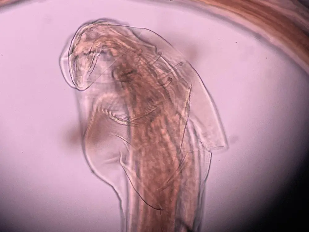
Diagnostic anatomy is compared with taxonomic descriptions, keys, and appropriate reference specimens. Calibrated lengths, widths, angles, and ratios are recorded where the structures and views support them. Hook geometry and other attachment structures are a focused application within HookFuse.
Measurements retain their units, calibration source, view, and reviewer. Missing calibration prevents claims about absolute physical size. Incomplete anatomy or conflicting metadata limits the taxonomic conclusion.
The proposed extension uses anatomically corresponding landmarks and reviewed semilandmarks to compare shape. Generalized Procrustes alignment removes differences in position, rotation, and overall scale; size is retained separately so allometry can be investigated.
Principal component analysis can describe shape variation without establishing species identity. Elliptic Fourier analysis is an optional branch for suitable closed outlines. View, orientation, preservation, and tracing error must be controlled before comparisons are interpreted.
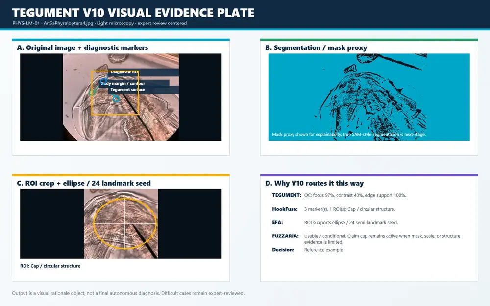
TEGUMENT organizes conservative image preparation, segmentation, and diagnostic-region review. Masks must preserve the anatomy needed for identification and measurement. Original photographs remain available alongside every processed derivative.
Vision Transformers are a proposed computer-vision extension for learning image features and testing candidate classification or reference-image comparison. Attention maps alone do not establish an anatomical explanation. Model version, reference data, preprocessing, and review status accompany any experimental output.
FUZZARIA organizes evidence adequacy and uncertainty to prioritize review. The proposed integration retains separate assessments for image quality, diagnostic coverage, calibration, and provenance, with explicit notes when analyses disagree.
Insufficient evidence can lead to recapture, further examination, or an unresolved record. No-detection images and inadequate acquisitions are retained with distinct outcomes. A model detecting no target is not, by itself, evidence that a host is uninfected.
The pilot compares conventional morphology and measurements with the added value of geometric morphometrics, Vision Transformers, and their combination. Expert-reviewed references are required before performance is reported.
Repeated images of a specimen remain in one data partition. Host and collection grouping are respected where required by the study design.
Detection, measurement error, shape repeatability, and taxonomic classification are evaluated separately. Unresolved cases remain visible.
FieldView documents acquisition, TEGUMENT prepares visual evidence, HookFuse organizes diagnostic characters, and FUZZARIA supports review.
Image preview only. No record is saved and no file is uploaded. Use the documentation template to prepare a linked record.
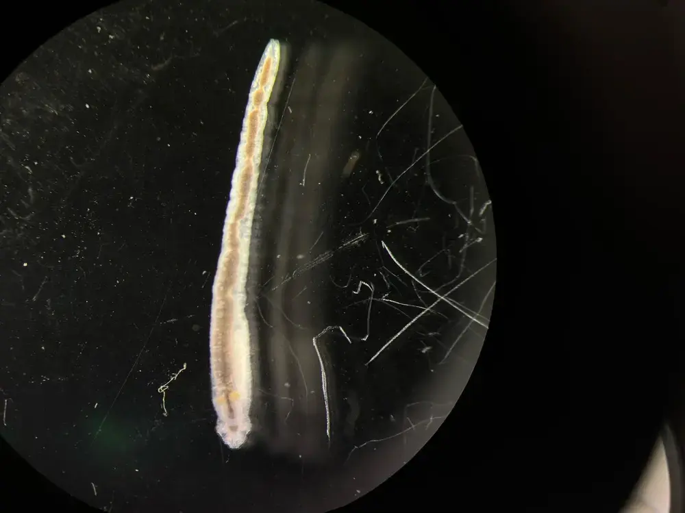
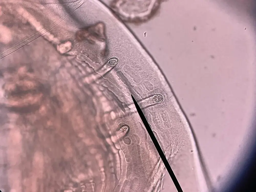
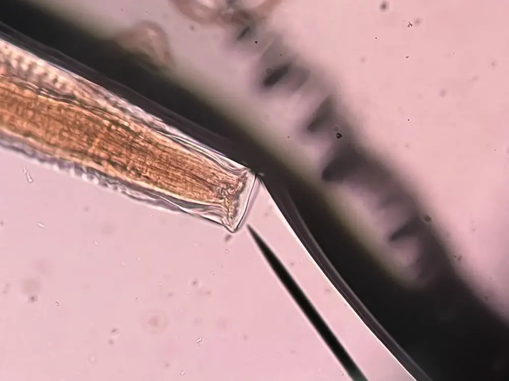
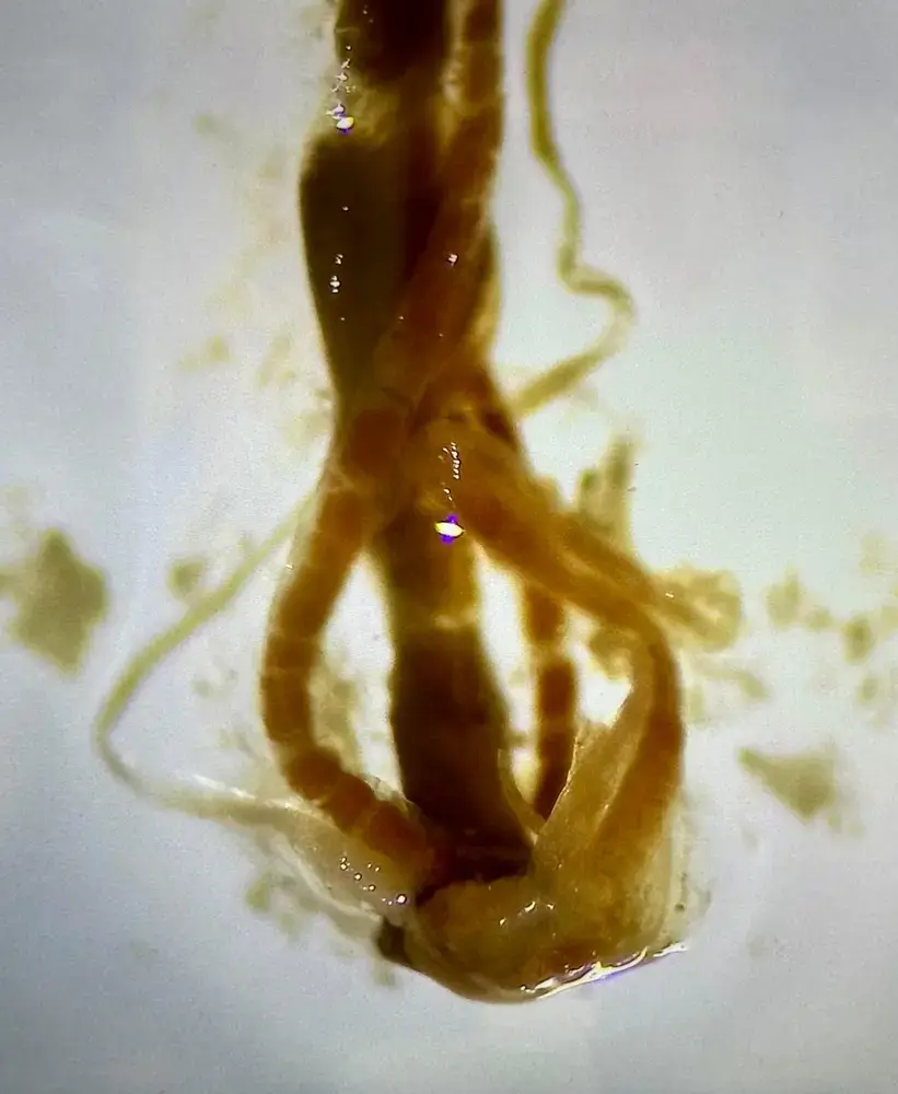
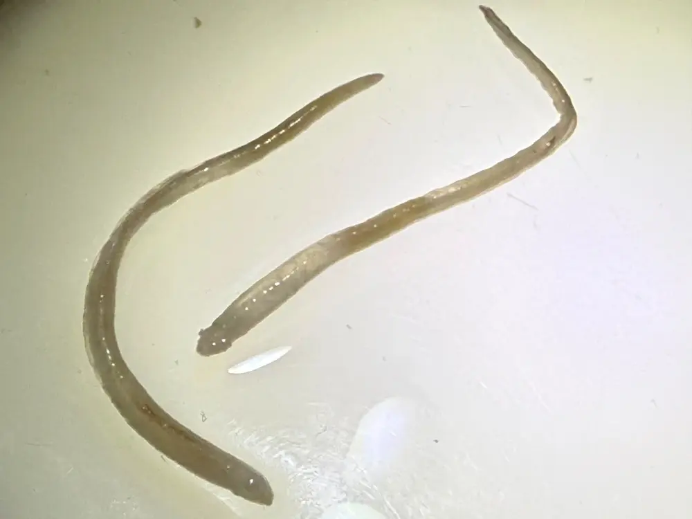
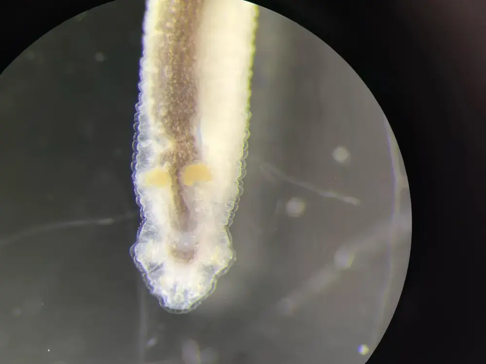
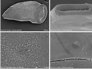
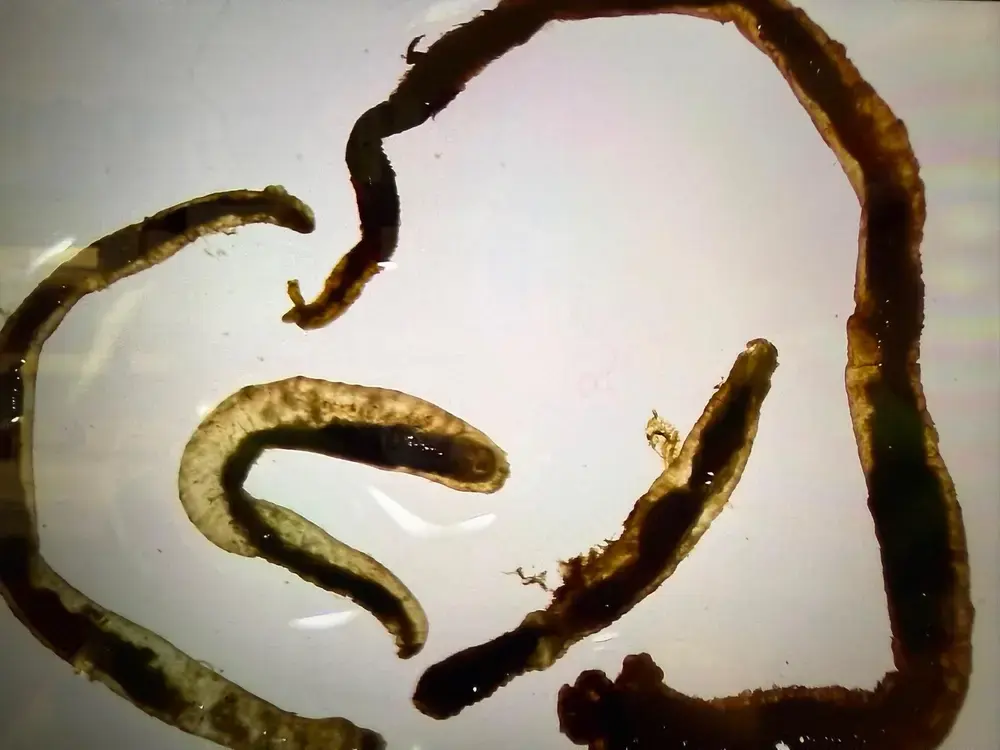
Original views, diagnostic details, and linked documentation.
These are image entries. Physical specimen identity, host association, and voucher linkage require verification. R-001, R-006, and R-017 now open structured draft records; their missing fields are shown explicitly.
A formal SEPS publication index will appear here as papers, natural history notes, technical reports and conference materials are released.

Society for Evolutionary Parasite Systematics
SEPS is a U.S. 501(c)(3) nonprofit research organization focused on parasite systematics and disease ecology. Comparative morphology, taxonomy, ecology, and preserved specimens guide our work on neglected and understudied parasites. Computer vision and quantitative shape analysis support these scientific questions.

For research partnerships, specialist review, and questions about specimens or images, contact M.R. Dwyer, Director of Research & Development.
Email SEPSSociety for Evolutionary Parasite Systematics